Proteins and their Structure

Each of the thousands of naturally occurring proteins has its own characteristic amino acid composition and sequence that result in a unique three-dimensional shape. Since the 1950s, scientists have determined the amino acid sequences and three-dimensional structures of numerous proteins and thus obtained important clues on how each protein performs its specific function in the body.
Proteins are large compounds that consist of chains of amino acids. Because of their great complexity, protein molecules cannot be classified on the basis of specific structural similarities, as carbohydrates and lipids are categorized. The two major structural classifications of proteins are based on far more general qualities: whether the protein is (1) fiberlike and insoluble or (2) globular and soluble.
Some proteins, such as those that compose hair, skin, muscles, and connective tissue, are fiberlike. These fibrous proteins are insoluble in water and usually serve structural, connective, and protective functions. Much of animal protein contains myosins, which are muscle proteins.
Globular proteins, the other major class, are soluble in aqueous media. In these proteins, the chains are folded so that the molecule as a whole is roughly spherical. Familiar examples include egg albumin from egg whites and hemoglobin in blood. Hemoglobin is important for transporting oxygen in the blood and gives muscle its red color.

Levels of Protein Structure

The structure of proteins is generally described as having four organizational levels. The first of these is the primary structure, which is the number and sequence of amino acids in a protein’s polypeptide chain. The primary structure of insulin, composed of 51 amino acids, is shown in below:
Two amino acid chains form the protein insulin. Chain A contains 21 amino acids, chain B contains 30 amino acids. The two chains are connected by disulfide bonds.
Primary Structure of Human Insulin. Human insulin, whose amino acid sequence is shown here, is a hormone that is required for the proper metabolism of glucose.

The primary structures of proteins are quite sturdy. In general, fairly vigorous conditions are needed to hydrolyze peptide bonds. The enzyme pepsin, which aids in the hydrolysis of proteins, is found in the digestive juices of the stomach (Greek pepsis, meaning “digestion”). Papain, another enzyme that hydrolyzes protein (in fact, it is used in meat tenderizers), is isolated from papayas.

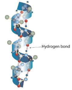
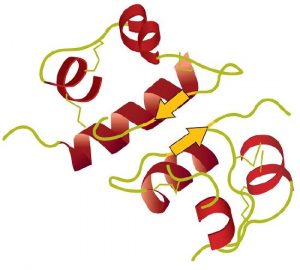
A protein molecule is not a random tangle of polypeptide chains. Instead, the chains are arranged in unique but specific 3D structures. The term secondary structure refers to the fixed arrangement of the polypeptide backbone. On the basis of X ray studies, Linus Pauling and Robert Corey postulated that certain proteins or portions of proteins twist into a spiral or a helix. This helix is stabilized by intrachain hydrogen bonding between two amino acids and is known as a right-handed α-helix. X ray data indicate that this helix makes one turn for every 3.6 amino acids, and the side chains of these amino acids project outward from the coiled backbone. The α-keratins, found in hair and wool, are exclusively α-helical in conformation. Some proteins have little or no helical structure, such as chymotrypsin, a digestive protein that breaks down other proteins. Others, such as hemoglobin, are helical in certain regions but not in others.

A Ball-and-Stick Model of an α-Helix. This ball-and-stick model shows the intrachain hydrogen bonding between amino acids. Each turn of the helix spans 3.6 amino acids. Note that the side chains (represented as green spheres) point out from the helix.
Another common type of secondary structure, called the β-pleated sheet conformation, is a sheetlike arrangement in which two or more extended polypeptide chains (or separate regions on the same chain) are aligned side by side. The aligned segments are connected by interchain hydrogen bonding. The β-pleated sheet is particularly important in structural proteins, such as silk fibroin. It is also seen in portions of many enzymes, such as lysozyme, an antibacterial protein found in egg whites.
A Ball-and-Stick Model of the β-Pleated Sheet Structure in Proteins. The side chains extend above or below the sheet and alternate along the chain. The protein chains are held together by interchain hydrogen bonding.
Tertiary structure refers to the unique three-dimensional shape of the protein as a whole, which results from the folding and bending of the protein backbone. The tertiary structure is intimately tied to the proper functioning of the protein. The figure below shows a depiction of the three-dimensional structure of insulin.
A Ribbon Model of the Three-Dimensional Structure of Insulin. The spiral regions represent sections of the polypeptide chain that have an α-helical structure, while the broad arrows represent β-pleated sheet structures.
Four major types of attractive interactions determine the shape and stability of the tertiary structure of proteins:
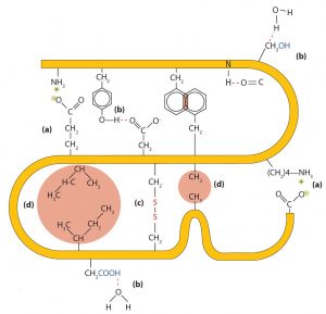
  1. Ionic bonding. Ionic bonds result from electrostatic attractions between positively and negatively charged side chains of amino acids. For example, the mutual attraction between a negative aspartic acid ion and a positive lysine ion helps to maintain a particular folded area of a protein (part (a) of the figure below).
  2. Hydrogen bonding. Hydrogen bonding forms between a highly electronegative oxygen atom or a nitrogen atom and a hydrogen atom attached to another oxygen atom or a nitrogen atom, such as those found in polar amino acid side chains. Hydrogen bonding (as well as ionic attractions) is extremely important in both the intra- and intermolecular interactions of proteins (part (b) of the figure below).
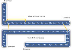
  3. Disulfide linkages. Two cysteine amino acid units may be brought close together as the protein molecule folds. Subsequent oxidation and linkage of the sulfur atoms in the two cysteine results in a disulfide linkage (part (c) of the figure below). Intrachain disulfide linkages are found in many proteins, including insulin (yellow bars in the structure of insulin above) and have a strong stabilizing effect on the tertiary structure.
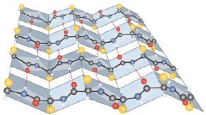
  4. Dispersion forces. Dispersion forces arise when a normally nonpolar atom becomes momentarily polar due to an uneven distribution of electrons, leading to an instantaneous dipole that induces a shift of electrons in a neighboring nonpolar atom. Dispersion forces are weak but can be important when other types of interactions are either missing or minimal (part (d) of the figure below). This is the case with fibroin, the major protein in silk, in which a high proportion of amino acids in the protein have nonpolar side chains. Because nonpolar groups cannot engage in hydrogen bonding, the protein folds in such a way that these groups are buried in the interior part of the protein structure, minimizing their contact with water.
Tertiary Protein Structure Interactions. Four interactions stabilize the tertiary structure of a protein: (a) ionic bonding, (b) hydrogen bonding, (c) disulfide linkages, and (d) dispersion forces.
When a protein contains more than one polypeptide chain, each chain is called a subunit. The arrangement of multiple subunits represents a fourth level of structure, the quaternary structure of a protein. Hemoglobin, with four polypeptide chains or subunits, is the most frequently cited example of a protein having quaternary structure (see figure below). The quaternary structure of a protein is produced and stabilized by the same kinds of interactions that produce and maintain the tertiary structure. A schematic representation of the four levels of protein structure is shown below:
The Quaternary Structure of Hemoglobin. Hemoglobin is a protein that transports oxygen throughout the body.
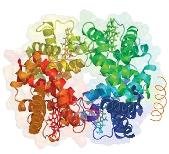
Source: Image from the RCSB PDB (www.pdb.org) of PDB ID 1I3D (R.D. Kidd, H.M. Baker, A.J. Mathews, T. Brittain, E.N. Baker (2001) Oligomerization and ligand binding in a homotetrameric hemoglobin: two high-resolution crystal structures of hemoglobin Bart’s (gamma(4)), a marker for alpha-thalassemia. Protein Sci. 1739–1749).

Summary of Levels of Protein Structure

Levels of Structure in Proteins

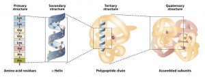
The primary structure consists of the specific amino acid sequence. The resulting peptide chain can twist into an α-helix or β-pleated sheet, which is one type of secondary structure. This helical segment is incorporated into the tertiary structure of the folded polypeptide chain. The single polypeptide chain is a subunit that constitutes the quaternary structure of a protein, such as hemoglobin that has four polypeptide chains.

Concept Review Exercises

  1. What is the predominant attractive force that stabilizes the formation of secondary structure in proteins?
  2. Distinguish between the tertiary and quaternary levels of protein structure.
  3. What name is given to the predominant secondary structure found in silk?
  4. What name is given to the predominant secondary structure found in wool protein?

Solutions

  1. hydrogen bonding
  2. Tertiary structure refers to the unique three-dimensional shape of a single polypeptide chain, while quaternary structure describes the interaction between multiple polypeptide chains for proteins that have more than one polypeptide chain.
  3. α-helix
  4. β-pleated sheet

Attributions

This page is based on “Chemistry 2e” by Paul Flowers, Klaus Theopold, Richard Langley, William R. Robinson, PhDOpenstax which is licensed under CC BY 4.0. Access for free at https://openstax.org/books/chemistry-2e/pages/1-introduction

This page is based on “The Basics of General, Organic, and Biological Chemistry” by David W Ball, John W Hill, Rhonda J ScottSaylor which is licensed under CC BY-NC-SA 4.0. Access for free at http://saylordotorg.github.io/text_the-basics-of-general-organic-and-biological-chemistry/index.html

License

Icon for the Creative Commons Attribution-NonCommercial-ShareAlike 4.0 International License

Chemistry of Food and Cooking Copyright © 2022 by Jessica Wittman is licensed under a Creative Commons Attribution-NonCommercial-ShareAlike 4.0 International License, except where otherwise noted.