9 Identify Anatomical Locations
Amanda Shelton
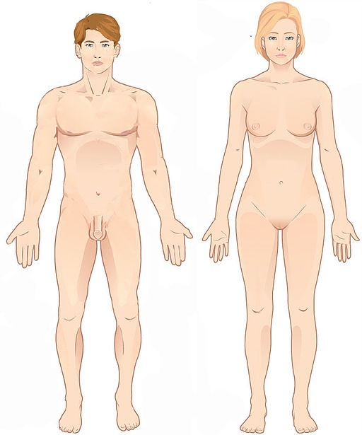
Anatomical Position
When it comes to describing movement and body areas, it is important to be able to identify anatomical locations. Whenever discussing the body and its position in relation to itself or others, it is important that we first identify the anatomical position. This position helps us to minimize confusion when referring to the human body by standardizing the body’s position.
“File:Anatomical position.jpg” by Connexions is licensed under CC BY 3.0
Description: a person standing facing forward with arms down at their side with their palms facing forward.
Describing Locations
Whenever we reference the body to describe locations, we will always use descriptions that compare body areas in relation to the anatomical position. We have several opposing descriptions that we can use to identify locations:
- right and left
- superior and inferior
- proximal and distal
- anterior and posterior
- medial and lateral
- internal and external
- contralateral and ipsilateral
- prone and supine
Right vs. Left
Most of the time, people are good about being able to identify their own right and left sides. It is important to consider that when we are describing anatomical locations that we always refer to the person’s left or right and not our own. This means that even if we are looking at the person (and they are looking at us) our left will be their right and our left will be their right. This can sometimes be confusing, especially when you are used to only describing your own right or left. For this reason, when we begin to discuss different movements later on in the chapter and throughout the textbook, sometimes it will be easier to use different and more specific descriptors than left or right.
Superior vs. Inferior
Superior and inferior descriptors refer to being above or below the reference point. Superior (or cranial) means that you are closer to the head than the reference point while inferior (or caudal) means that you are closer to the feet than the reference point. Remember – we are always describing locations in reference to the anatomical position, so even if someone changes their body position the description of locations using superior/inferior would remain the same.
Superior/Inferior Examples
The knee joint is inferior to the hip joint.
The sternum is superior to the pelvis.
The lumbar vertebrae are inferior to the cervical vertebrae.
The carpals are superior to the phalanges.
Proximal vs. Distal
Proximal and distal descriptors refer to being further or nearer to the center of the body or the point of reference being discussed. Proximal means that you are nearer to the center of the body or reference point while distal means you are further from the center of the body or reference point. Again, when we reference the center of the body, this is in relation to the anatomical position.
Proximal/Distal Examples
The distal end of the humerus is at the elbow.
The hip joint is proximal to the ankle joint.
The nasal bone is distal to the mandible.
The proximal end of the femur (the femoral head) articulates with the pelvis to create the hip joint.
Anterior vs. Posterior
Anterior and posterior refer to being on or toward the front or back or the body, with the exception of the feet. Anterior (or ventral) means that you are on or toward the front or palm side of the body while posterior (or dorsal) means that you are on or toward the back side of the body. The exception to the rule of these terms as descriptors are your feet where the dorsal aspect is the top of your foot and the ventral aspect is the bottom of your foot. With this exception, one easy way to remember because the palm side of your hands are anterior and that is matched on the bottom of your feet. We also see the terms anterior and posterior descriptors used in some of our naming with various soft tissue components in the body such as the tibialis posterior, a muscle in the distal low leg crossing through the ankle joint to help with movements like plantar flexion and inversion of the ankle, which we will discuss later on.
Anterior/Posterior Examples
The patella is on the anterior aspect of your knee.
A triceps extension activates the posterior musculature of the upper arm.
Medial vs. Lateral
Medial and lateral refer to the position in relation to the midline of the body. We can visualize the midline by pretending that we are holding a string from the top of our heads and positioning that string so that is falls straight down our body directly between our eyes. That string’s position represents the midline of the body. This means that when we look at something like the arms or legs, we can refer to the medial and lateral aspects of those body parts in relation to where the ‘string’ is. Medial means that you are closer to the midline of the body while lateral means that you are further from the midline of the body.
Medial/Lateral Examples
We see medial and lateral represented within the naming of some of the soft tissue components of our body like the medial collateral ligament (MCL) and the lateral collateral ligament (LCL) in our knees.
- Medial Collateral Ligament of the knee is on the side of the knee closer to the midline (“inside” aspect of the knee).
- Lateral Collateral Ligament of the knee is on the side of the knee further from the midline (“outside” aspect of the knee).
Radial vs. Ulnar
We also see this being described similarly in other areas of the body using more localized terms but for a similar purpose. In the arm, we use the radius and ulna, the two bones in the forearm to describe a similar comparison as medial and lateral. In the anatomical position, the ulna is on the medial aspect of the forearm (pinky side/ulnar side) and the radius is on the lateral aspect of the forearm (thumb side/radial side). With this, we will have different movements and components of anatomy that will use radial and ulnar in place of medial and later.
Radial/Ulnar Examples
- In the elbow we have the ulnar collateral ligament (UCL) on the medial aspect of the joint
- When we go into the movement you think might be called lateral flexion of the wrist, we instead refer to it as radial deviation.
Internal vs. External
We also have the descriptors internal and external. In a similar way to medial and lateral referring to position in relation to the midline of the body, we can also use the terms internal and external. We can have internal rotation where we are rotating toward the midline of the body and external rotation where we are rotating away from the midline of the body. We will discuss internal and external rotation more when we discuss muscle actions and movements. We can use internal and external to describe being located more inside the body (internal) vs. outside the body (external).
Contralateral vs. Ipsilateral
The last of the descriptors that we will discuss is contralateral and ipsilateral. These terms will reference areas as being on the same side or opposite side as they compare to a reference point. Contralateral would be used to describe something on the opposite side of the body from the reference point whereas ipsilateral would be used to describe something on the same side of the body as the reference point.
Contralateral/Ipsilateral Examples
If we are referring to a right ankle:
- their gastrocnemius on the ipsilateral leg is the calf muscle on their right side.
- their knee on the contralateral leg would be their left knee.
Prone vs. Supine
A body that is lying down is described as either prone or supine. Prone describes a face-down orientation, and supine describes a face up orientation. These terms are sometimes used in describing the position of the body during specific physical exercises. This can also be used to describe hand placement – you can think of supine as being able to hold soup in your hand when your palms are face up. When your palms are facing down, you can’t hold soup in your hand so you know it is in the prone position!
The reference position for the body used when describing positions and directions. The person is facing forward with arms at their side, palms facing forward and toes pointed forward.
anatomical directional descriptor for closer to the head than the reference point
anatomical directional descriptor for closer to the feet/toes than the reference point
anatomical directional descriptor for nearer to the center of the body or reference point
anatomical directional descriptor for further from the center of the body than the reference point
anatomical directional descriptor for on or toward the front or palm side of the body
anatomical directional descriptor for on or toward the back side of the body
anatomical directional descriptor for closer to the midline of the body
anatomical directional descriptor for further from the midline of the body
anatomical directional descriptor for more inside the body or further beneath the surface of the body
anatomical directional descriptor for more outside the body or closer to the surface of the body
anatomical directional descriptor for the opposite side of the body from the reference point
anatomical directional description for something on the same side of the body as the reference point
anatomical directional descriptor for something face down or palm down
anatomical directional descriptor for face up or palm up orientation
