Chapter 3 – Cardiorespiratory Fitness
Cardiovascular Anatomy
The cardiorespiratory system operates to obtain and circulate vital compounds throughout the body—specifically, oxygen and nutrients, such as food energy, vitamins, and minerals. Both oxygen and nutrients, which are imperative for cellular energy production, must be taken in from the lungs and digestive system. Because the heart and lungs are so interlocked in this process, the two systems are often labeled together as the cardiorespiratory system. Without a healthy respiratory system, the body would struggle to bring in enough oxygen, release carbon dioxide (the chemical waste product of cellular metabolism) and eliminate unwanted particles that enter the respiratory tract when inhaling. Without a healthy heart, transporting oxygen from the lungs and nutrients from the digestive system to the body’s cells would be impossible. If the health of the cardiorespiratory system were compromised enough, survival would be impossible.
The cardiovascular system is comprised of the heart and blood vessels of the body. These structures function primarily to provide circulate oxygenated blood to and remove waste from body tissues.
Shape and Size of the Heart
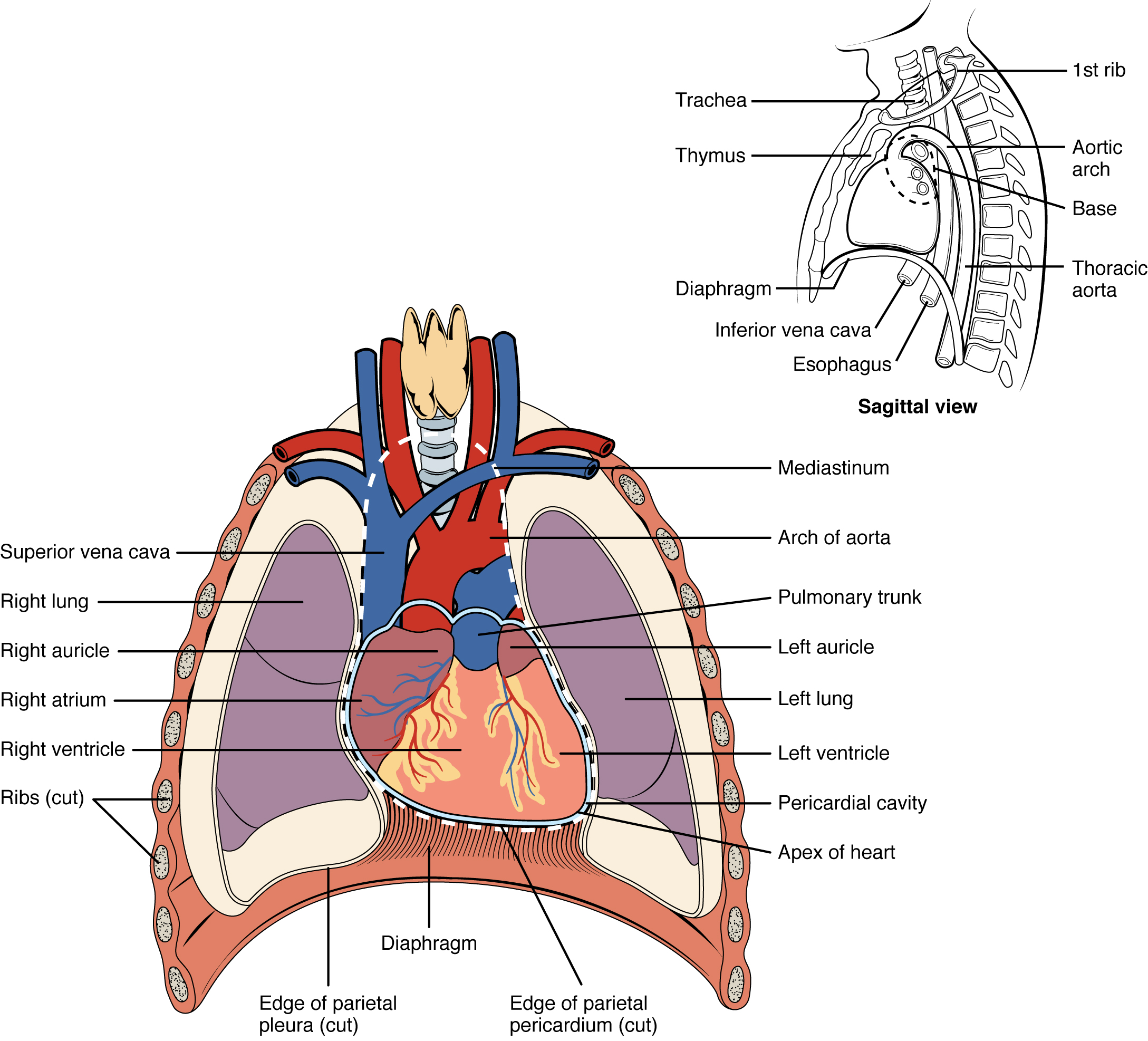
A typical heart is approximately the size of your fist: 12 cm (5 in) in length, 8 cm (3.5 in) wide, and 6 cm (2.5 in) in thickness. Given the size difference between most members of the sexes, the weight of a female heart is approximately 250–300 grams (9 to 11 ounces), and the weight of a male heart is approximately 300–350 grams (11 to 12 ounces). The heart of a well-trained athlete, especially one specializing in aerobic sports, can be considerably larger than this. Cardiac muscle responds to exercise in a manner similar to that of skeletal muscle. That is, exercise results in the addition of protein myofilaments that increase the size of the individual cells without increasing their numbers, a concept called hypertrophy. Hearts of athletes can pump blood more effectively at lower rates than those of nonathletes.
Chambers and Circulation through the Heart
The human heart consists of four chambers: The left side and the right side each have one atrium and one ventricle. Each of the upper chambers, the right atrium and the left atrium, acts as a receiving chamber and contracts to push blood into the lower chambers, the right ventricle and the left ventricle. The ventricles serve as the primary pumping chambers of the heart, propelling blood to the lungs or to the rest of the body.
There are two distinct but linked circuits in the human circulation called the pulmonary and systemic circuits. Although both circuits transport blood and everything it carries, we can initially view the circuits from the point of view of gases. The pulmonary circuit transports blood to and from the lungs, where it picks up oxygen and delivers carbon dioxide for exhalation. The systemic circuit transports oxygenated blood to virtually all of the tissues of the body and returns relatively deoxygenated blood and carbon dioxide to the heart to be sent back to the pulmonary circulation.
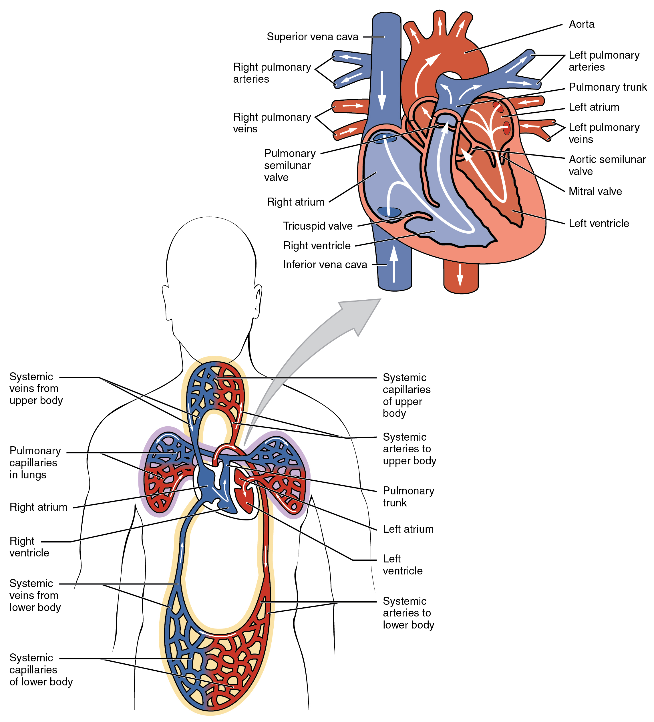
Dual System of the Human Blood Circulation:
Blood flows from the right atrium to the right ventricle, where it is pumped into the pulmonary circuit. The blood in the pulmonary artery branches is low in oxygen but relatively high in carbon dioxide.
Gas exchange occurs in the pulmonary capillaries (oxygen into the blood, carbon dioxide out), and blood high in oxygen and low in carbon dioxide is returned to the left atrium.
From here, blood enters the left ventricle, which pumps it into the systemic circuit. Following exchange in the systemic capillaries (oxygen and nutrients out of the capillaries and carbon dioxide and wastes in), blood returns to the right atrium and the cycle is repeated.
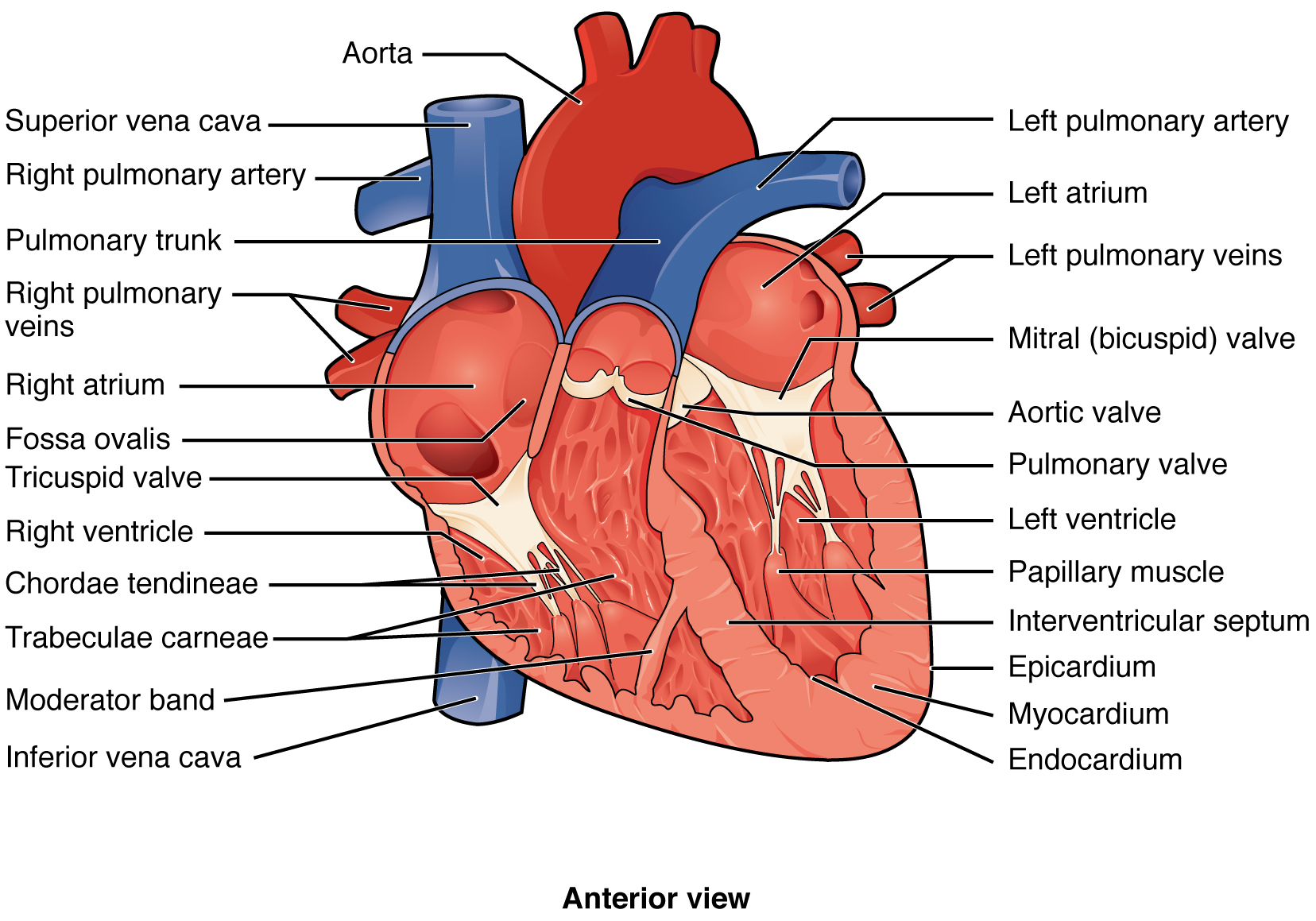
Internal Structures of the Heart:
This anterior view of the heart shows the four chambers, the major vessels and their early branches, as well as the valves. The presence of the pulmonary trunk and aorta covers the interatrial septum, and the atrioventricular septum is cut away to show the atrioventricular valves.
